
Page 322 - SAHCS HIVMed Journal Vol 20 No 1 2019
P. 322
Page 4 of 6 Original Research
TABLE 4: Normal values for Inkosi Albert Luthuli Central Hospital Electrophysiology Laboratory.
Nerve Distal motor latency (ms) Amplitude NCV (m/s) F latency (ms)
Peroneal nerve (EDB) < 6.5 > 4mV > 41 < 57
Tibial nerve ( AHB) < 5.9 > 4mV > 40 < 57
Ulnar nerve (ADM) < 3.6 > 6mV > 51 < 32
Median nerve (APB) < 4.5 > 4mV > 48 < 33
Sural (Stimulation site = 14cm) < 4.5 > 7uV - -
Superficial peroneal (Stimulation site = 14cm) < 3.8 > 7uV - -
Ulnar SNAP < 2.1 > 10uV - -
Median SNAP < 2.3 > 15uV - -
Source: In-house collaboration among neurophysiologists from Pretoria Academic and Groote Schuur Hospital, SA Neurology Association Meeting, Rustenburg, 1998
EBD, Extensor Digitorium Brevis; AHB, Abductor Hullucis Brevis; ADM, Adductor Digiti Minimi; APB, Abductor Pollicis Brevis; SNAP, Sensory Nerve Action Potential; ms, millisecond; NCV, nerve
conduction velocity; m/s, metre per second.
TABLE 5: Needle Examination.
Muscle Spontaneous activity Motor unit potential Recruitment pattern
Insertional activity Fib PSW Fasic Amplitude Duration Polyphasia
Lumbar paraspinals 2+ (3+ in patient 11, 7) 3+ 2+ 0 normal normal 3+ Reduced (Single unit recruitment in patient 11)
Gluteus medius 1+ 2+ 1+ 0 normal normal 2+ Reduced (Single unit in patient 1,9,11)
Quadriceps 1+ 1+ 1+ 0 normal normal 2+ Reduced (Single unit recruitment in patient 11)
Tibialis anterior 1+ 1+ 1+ 0 normal normal 1+ Reduced
Gastrocnemius 1+ 1+ 1+ 0 normal normal 1+ Reduced
Fib, Fibrillation potentials; PSW, Positive sharp waves; Fasic, Fasiculations.
therapy by 4 months had minimal residual deficit at
a b 18 months follow-up with a mRS of 1. The median time for
recovery in all categories was 3.4 months (IQR 1.8–5.6).
There were no relapses during the 18-month follow-up.
Within the period of corticosteroid therapy, there were
no documented side effects and no patients required
corticosteroid sparing immunosuppressive agents or long-
term corticosteroids therapy. Six patients had CD4 counts
< 350 cells/µL and qualified for ART according to ART
guidelines at that time. HIV titres were not documented.
Three patients were commenced on ART at 4 months after
the diagnosis. These three patients had recovered prior
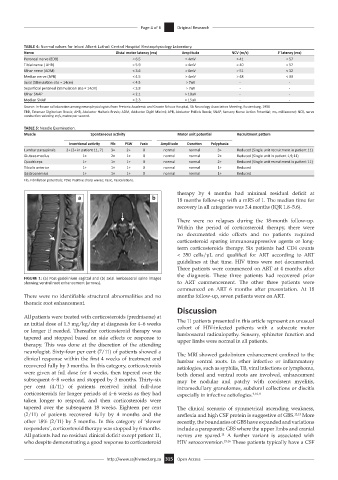
FIGURE 1: (a) Post-gadolinium sagittal and (b) axial lumbosacral spine images
showing ventral root enhancement (arrows). to ART commencement. The other three patients were
commenced on ART 6 months after presentation. At 18
There were no identifiable structural abnormalities and no months follow-up, seven patients were on ART.
thoracic root enhancement.
Discussion
All patients were treated with corticosteroids (prednisone) at
an initial dose of 1.5 mg/kg/day at diagnosis for 4–6 weeks The 11 patients presented in this article represent an unusual
or longer if needed. Thereafter corticosteroid therapy was cohort of HIV-infected patients with a subacute motor
tapered and stopped based on side effects or response to lumbosacral radiculopathy. Sensory, sphincter function and
upper limbs were normal in all patients.
therapy. This was done at the discretion of the attending
neurologist. Sixty-four per cent (7/11) of patients showed a The MRI showed gadolinium enhancement confined to the
clinical response within the first 4 weeks of treatment and lumbar ventral roots. In other infective or inflammatory
recovered fully by 3 months. In this category, corticosteroids aetiologies, such as syphilis, TB, viral infections or lymphoma,
were given at full dose for 4 weeks, then tapered over the both dorsal and ventral roots are involved, enhancement
subsequent 6–8 weeks and stopped by 3 months. Thirty-six may be nodular and patchy with coexistent myelitis,
per cent (4/11) of patients received initial full-dose intramedullary granulomas, subdural collections or discitis
corticosteroids for longer periods of 4–6 weeks as they had especially in infective aetiologies. 9,10,11
taken longer to respond, and then corticosteroids were
tapered over the subsequent 18 weeks. Eighteen per cent The clinical scenario of symmetrical ascending weakness,
(2/11) of patients recovered fully by 4 months and the areflexia and high CSF protein is suggestive of GBS. 12,13 More
other 18% (2/11) by 5 months. In this category of ‘slower recently, the boundaries of GBS have expanded and variations
responders’, corticosteroid therapy was stopped by 6 months. include a paraparetic GBS where the upper limbs and cranial
14
All patients had no residual clinical deficit except patient 11, nerves are spared. A further variant is associated with
who despite demonstrating a good response to corticosteroid HIV seroconversion. 15,16 These patients typically have a CSF
http://www.sajhivmed.org.za 315 Open Access