
Page 321 - SAHCS HIVMed Journal Vol 20 No 1 2019
P. 321
Page 3 of 6 Original Research
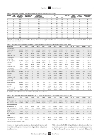
TABLE 1: Demographic, laboratory, and radiological features of motor lumbosacral radiculopathy.
Number Age CD4 count mRS Scores at Duration of CSF MRI (RE) Time to mRS at Relapses within
(Years) at diagnosis presentation progression of recovery 18 months 18 months
(cells/μL) symptoms (Months) P L Glu Prot (Months)
1. 27 657 4 3.0 0 32 3.2 0.98 Y 3 0 Nil
2. 22 155 4 2.0 0 0 3.7 1.02 Y 2 0 Nil
3. 42 480 5 4.0 0 16 2.3 3.68 Y 3 0 Nil
4. 29 149 5 3.5 0 4 3.1 1.83 Y 4 0 Nil
5. 21 265 5 2.0 2 17 3.3 1.02 Y 5 0 Nil
6. 24 450 4 4.0 1 18 2.8 1.69 Y 4 0 Nil
7. 18 380 4 4.0 0 24 3.6 2.61 Y 3 0 Nil
8. 51 140 4 2.0 0 0 3.4 0.77 y 2 0 Nil
9. 32 124 4 5.0 1 12 4.2 1.52 Y 3 0 Nil
10. 40 112 5 4.0 2 9 4.1 0.89 Y 5 0 Nil
11. 43 389 4 2.0 0 5 3.2 1.34 Y 4 1 Nil
mRS, Modified Rankin Scale; CSF, cerebrospinal fluid; MRI, magnetic resonance imaging; RE, root enhancement; P, polymorphocyte count (cells/μL); L, lymphocyte count (cells/μL); Glu, glucose
(mmol/L); Prot, protein (g/dL); Y, yes.
TABLE 2: Electrophysiological findings of patients with motor lumbosacral radiculopathy in HIV-infected patients: Motor studies.
Motor nerve Pat: 1 Pat: 2 Pat: 3 Pat: 4 Pat: 5 Pat: 6 Pat: 7 Pat: 8 Pat: 9 Pat: 10 Pat: 11 Median IQR
Peroneal ( R/L)
Amplitude (mV) 2.2/2.4 4.2/3.8 1.9/2.3 4.4/3.8 2.6/1.8 1.8/2.6 4.1/4.4 5.2/4.8 0.9/1.1 3.4/3.6 5.2/5.1 3.6 2.2–4.3
DML (ms) 5.2/4.8 4.8/5.1 6.2/6.1 5.9/5.8 5.4/5.6 6.3/6.2 5.8/5.9 5.5/4.9 6.9/7.1 4.6/4.2 4.4/4.2 5.5 4.8–6.5
CV (m/s) 44/46 46/48 41/43 40/41 44/46 41/43 44/45 42/43 38/41 42/44 38/40 43 41–44
F Response (ms) 58/61 62/68 59/58 73/75 78/68 59/58 59.5/61 abs/64 abs/abs 62/67 73/75 62 59–70.5
F Estimate (ms) 51/50 49/50 52/53 58/54 51/53 52/53 54/55 48/49 abs/abs 57/58 61/62 53 50–55
Tibial ( R/L)
Amplitude (mV) 3.1/3.5 2.8/2.6 1.8/2.1 4.2/4.4 3.2/3.8 2.8/2.6 3.9/4.1 6.7/7.2 1.2/2.1 2.8/2.6 6.1/6.8 3.5 2.6–4.2
DML (ms) 7/6.6 5.8/5.6 6.8/6.6 4.8/4.9 5.9/6.1 5.8/5.6 4.3/4.5 5.3/5.5 6.8/6.9 6.8/6.6 5.9/6.1 3.5 2.6–4.2
CV (m/s) 48/49 40/41 42/44 44/45 51/48 41/40 45/46 43/42 40/39 41/40 43/44 45 43–46
F Response (ms) 62/65 70/68 64/74 64/62 65/68 69/70 abs/70 65/72 abs/71 69/70 64/62 68 64–70
F Estimate (ms) 52/52 54/56 48/49 54/55 51/52 54/56 55/abs 53/54 58/abs 54/56 56/54 54 52–55
Median ( R/L)
Amplitude (mV) 8.2/8.5 10.2/10 n/a n/a n/a 12/12.8 8.6/8.1 10.9/11.8 n/a 11.1/10.9 6.1/6.2 10.3 8.2–11
DML (ms) 3.2/3.3 4.1/4.2 n/a n/a n/a 4.1/3.8 3.9/3.8 4.4/4.5 n/a 4.1/4.2 3.9/3.8 4 3.8–4.2
CV (m/s) 58/52 58/56 n/a n/a n/a 55/56 48/45 44/45 n/a 51/50 49/51 51 48–56
F Response (ms) 28/29 31/29 n/a n/a n/a 24/28 24/26 28/32 n/a 31/33 29/28 29 27.5–31
F Estimate (ms) 31/30 30/31 n/a n/a n/a 28/28 32/30 33/34 n/a 35/32 32/34 31 30–31
Ulnar ( R/L)
Amplitude (mV) 6.9/6.3 5.8/5.2 n/a n/a n/a 8.8/9.1 7.9/6.1 9.8/9.1 n/a 8.6/9.1 9.4/9.2 8.8 6.6–9.1
DML (ms) 2.6/2.1 3.3/3.1 n/a n/a n/a 2.9/2.6 2.6/2.1 3.1/2.7 n/a 2.9/2.6 3.2/2.8 2.7 2.6–3
CV (m/s) 50/48 58/56 n/a n/a n/a 54/56 52/49 46/48 n/a 52/54 52/56 52 49–55
F Response (ms) 31/29 26/24 n/a n/a n/a 28/25.5 31/29 31/30 n/a 28/25.5 31/30 29 26–30
F Estimate (ms) 30/30 26/27 n/a n/a n/a 30/31 33/32 34/35 n/a 30/31 34/35 31 30–33
R/L, Right/Left; DML, distal motor latency; CV, conduction velocity; IQR, interquartile range.
TABLE 3: Electrophysiological findings in patients with motor lumbosacral radiculopathy in HIV-infected patients: Sensory studies.
Sensory nerves Pat:1 Pat:2 Pat:3 Pat:4 Pat:5 Pat:6 Pat:7 Pat:8 Pat:9 Pat: 10 Pat:11 Median IQR
Sural
Amplitude (µV) R/L 9.8/10.5 12.5/12.3 11.2/12.3 10.3/9.5 11.5/11.6 12.2/12.9 14.1/13.9 9.5/9.3 16/15.8 12.2/13.1 15.1/15 12.5 10–13
Peak latency (ms) R/L 4.1/4.3 3.8/4 4.2/4.1 3.9/4.1 4.2/3.8 4.4/4.2 3.9/4.1 3.9/3.8 4.4/4.5 2.9/3.1 3.9/3.6 4.1 3.9–4.2
Superficial Peroneal
Amplitude (µV) R/L 3.5/4 6.7/6 5.6/5 4.8/5 6.6/6 7.3/7.5 6.5/6.1 7.1/7.4 6.8/6.5 7.1/7.7 7.8/7 6.5 5.7–7.1
Peak latency (ms) R/L 3.1/3.3 2.9/2.8 2.6/2.8 3.2/3.3 3.8/3.6 2.1/2.5 2.2/2.6 3.2/3.8 3.1/2.9 3.5/3.3 3.2/3 3.1 2.27–3.3
Median
Amplitude µV R/L 45/58 68/62 n/a n/a n/a 85/76 58/56 115/98 n/a 45/48 85/96 65 56.5–85
Peak latency R/L 3.4/3.6 3.3/3.1 n/a n/a n/a 2.6/2.9 2.7/2.9 3.1/2.9 n/a 3.1/3.3 2.9/3.2 3 2.9–3
Ulnar
Amplitude µV R/L 24/28 45/44 n/a n/a n/a 48/53 77/76 65/64 n/a 32/38 38/27 44 33–61
Peak latency R/L 2.2/2 2.1/2.6 n/a n/a n/a 2.8/3 2.1/2.7 2.8/2.6 n/a 2.6/2.3 2.1/2.3 2 2.3–2.8
R/L, Right/Left; IQR, interquartile range.
reduced or single unit recruitment of polyphasic motor unit All 11 patients had MRI with gadolinium, of the thoracolumbar
potentials with greater involvement of proximal rather than and lumbosacral spine. Imaging revealed root enhancement
distal muscles. of the lumbosacral ventral roots in all patients (Figure 1).
http://www.sajhivmed.org.za 314 Open Access